Table of Contents
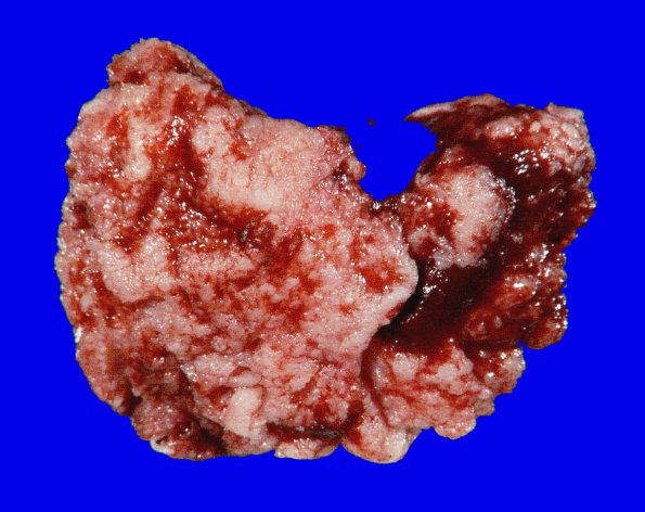
Washington University Experience | NEOPLASMS (MENINGIOMA) | Gross Pathology | 68A Meningioma, Atypical (Case 68) Gross_4
Case 68 History ---- The patient was a 58 year old woman who presented with mental status changes. MRI showed large left frontal parietal dural-based enhancing mass. Operative procedure: Left parietal craniotomy for tumor resection. ---- 68A The external and cut sections of this meningioma. ---- Sections of left parietal tumor reveal a focally hypercellular neoplasm arranged in whorls and fascicles with moderately sized cells containing oval nuclei, small nucleoli with and abundant eosinophilic cytoplasm. In some areas the tumor cells have macronucleoli. Focal areas show small cell features and sheeting of tumor cells. There are multiple areas of tumor necrosis. Mitotic figures are present, reaching up to 9/10 high power fields focally. ---- The morphological features are consistent with the diagnosis of atypical meningioma, WHO Grade II.

