Table of Contents
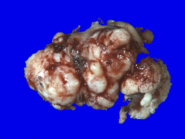
Washington University Experience | NEOPLASMS (MENINGIOMA) | Gross Pathology | 69A1 Meningioma, atypical (Case 69) 1
Case 69 History ---- The patient is a 70-year-old woman with history of headaches and declining cognitive status. Operative procedure: Resection. ---- Sections consist of a meningothelial neoplasm arranged mostly in a sheet-like pattern with focal areas of fascicular to nodular growth pattern. There is abundant collagen deposition within this tumor. The individual tumor cells are polygonal to spindle-shaped with ill-defined cytoplasmic membranes, eosinophilic cytoplasm and vesicular nuclei with focally prominent nucleoli. In some areas, the tumor is quite cellular. Scattered mitoses are identified throughout the tumor and in hypercellular areas the count reaches 7/10 HPF. Rare psammoma bodies are present. The tumor shows focal expression of progesterone receptor and a high Ki-67 labeling index, focally reaching up to 9.2%. ---- Comment: The overall findings are those of an atypical meningioma, WHO Grade II.

