Table of Contents
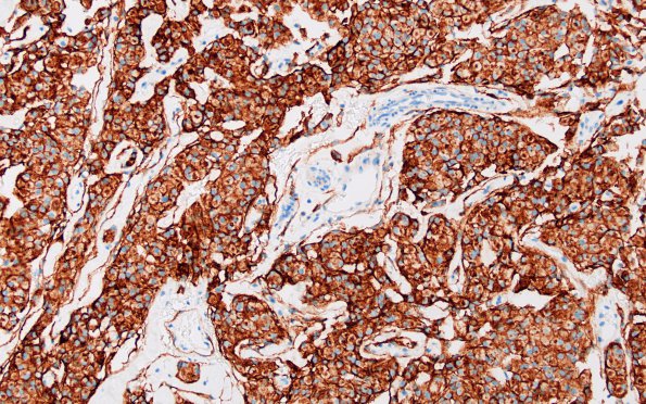
Washington University Experience | NEOPLASMS (MENINGIOMA) | Meningothelial | 1B Meningioma (Case 1) EMA 1
EMA staining is typically patchy and very membrane localized rather than the more homogenous pattern seen here (EMA IHC), probably contributed by a thick section.

