Table of Contents
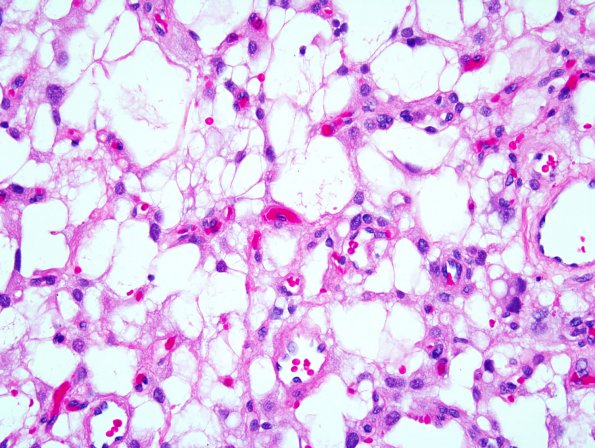
Washington University Experience | NEOPLASMS (MENINGIOMA) | Microcystic | 1A1 Meningioma, microcystic (Case 1) 3.jpg
Case 1 History ---- The patient was a 34 year old man who presented with a 3.5 cm right frontal dural-based mass. ---- 1A1-3 This is a low to moderately cellular neoplasm with prominent microcystic spaces and numerous hyalinized vessels. The majority of tumor cells are associated with thin wispy cytoplasmic processes creating a cobweblike growth pattern. However, rare epithelioid cells arranged in whorls are also encountered. Scattered bizarre or markedly atypical nuclei are also identified. However, there is no associated increase in mitotic activity or evidence of necrosis, hypercellularity, or macronucleoli. (H&E)

