Table of Contents
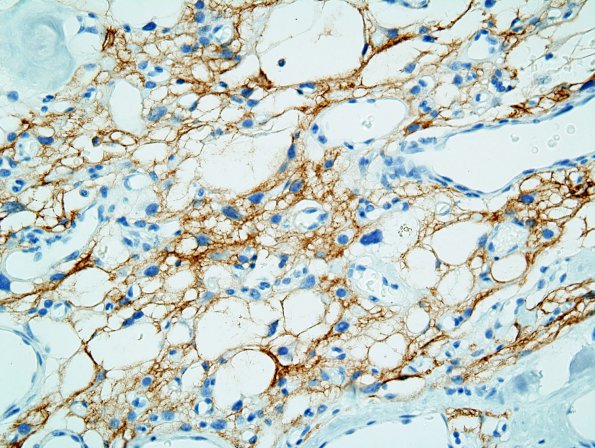
Washington University Experience | NEOPLASMS (MENINGIOMA) | Microcystic | 1B2 Meningioma, microcystic (Case 1) EMA 3.jpg
Tumor cells display convincing delicate immunoreactivity of processes for epithelial membrane antigen (EMA), associated with a patchy membrane-associated staining pattern.

