Table of Contents
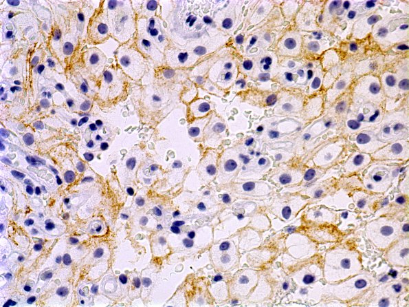
Washington University Experience | NEOPLASMS (MENINGIOMA) | Microcystic | 2B Meningioma, microcystic (Case 2) EMA
Immunohistochemical stains show that the tumor cells have patchy areas with crisp membranous staining for EMA. ---- Ancillary data (not shown): Tumor cells have scattered nuclear positivity for progesterone receptor and the MIB-1 labeling index is low. They are negative for S100 protein and inhibin. The histology and immunohistochemical staining pattern is consistent with that of a meningioma, microcystic variant, WHO grade I.

