Table of Contents
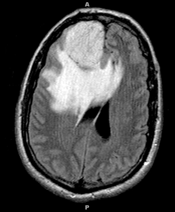
Washington University Experience | NEOPLASMS (MENINGIOMA) | Microcystic | 4A1 Meningioma, Microcystic Angiomatous (Case 4) MRI 3 - Copy
Case 4 History ---- The patient was a 57 year old man with a right frontal tumor. Operative procedure (9/10/09): ---- MRI studies show hyperintensity of the tumor and marked surrounding parenchymal edema as seen in this FLAIR scan.

