Table of Contents
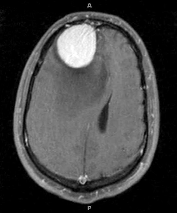
Washington University Experience | NEOPLASMS (MENINGIOMA) | Microcystic | 4A2 Meningioma, Microcystic Angiomatous (Case 4) MRI 1 - Copy
4A2,3 These T1-weighted images with administered contrast are shown in axial (4A2) and sagittal (4A3) scans.

