Table of Contents
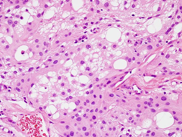
Washington University Experience | NEOPLASMS (MENINGIOMA) | Microcystic | 5A4 Meningioma, microcystic (Case 5) H&E 2
There is also abundant hyalinized stroma. In addition, areas of microcystic changes can be seen throughout. (H&E) ---- Ancillary findings (not shown): Immunostains demonstrate the tumor cells are patchy and weakly positive for progesterone receptor and EMA. Ki-67 is variable, with some increased hotspots; however, mitoses are infrequent. The morphology and immunophenotype are diagnostic of meningioma, WHO Grade 1, with microcystic features and increased proliferation indices.

