Table of Contents
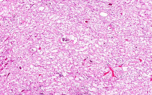
Washington University Experience | NEOPLASMS (MENINGIOMA) | Microcystic | 6A1 Meningioma, microcystic (Case 6) H&E 7
Case 6 History ---- The patient was a 68 year old woman with cerebral infarction with difficulty speaking. MRI brain revealed a 3.5 x 2.87 x 3.64 cm partially enhancing mass at the surface of the right parietal lobe with surrounding edema extending to the posterior horn of the right lateral ventricle. 6A1-3 H&E stained tumor shows a neoplasm composed of cells with round to oval, hyperchromatic nuclei, and clear to pale eosinophilic cytoplasm with microcystic changes. There is significant degenerative nuclear atypia, and occasional cells have prominent nucleoli. No obvious intranuclear clearing or pseudoinclusions are present. Many cells have thin, elongated processes surrounding cyst-like structures, some empty and other containing granular debris. Many areas of the tumor display numerous thin-walled vessels, which are often hyalinized. Occasional eosinophilic globules are present. Mitotic figures are present up to 4/10 HPF. Brain parenchyma is present on all three sections, but brain invasion is not identified.

