Table of Contents
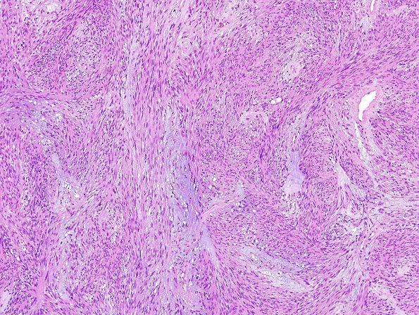
Washington University Experience | NEOPLASMS (MENINGIOMA) | Mucinous | 2A1 Meningioma, mucinous (Case 1) H&E 2.jpg
2A1-3 H&E stained sections of each of the four 2013 resection specimens show meningiomas with predominant chordoid morphology, characterized by fascicles of eosinophilic spindled cells with abundant basophilic stromal mucin and a vacuolated appearance, interlaced with more compact fascicles of eosinophilic spindled cells. Hyalinized blood vessels are frequent. The tumor cells have modest eosinophilic cytoplasm, oval moderately pleomorphic nuclei, and occasional prominent nucleoli. Mitotic figures are rare and do not exceed a density of 1/10HPF. There is no evidence of necrosis, sheeting (loss of architecture), small cell change, or hypercellularity. Brain tissue is associated with the tumor, but there is no evidence of brain invasion.

