Table of Contents
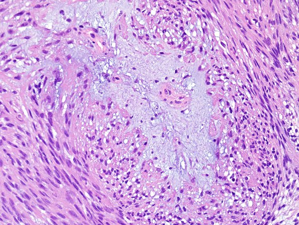
Washington University Experience | NEOPLASMS (MENINGIOMA) | Mucinous | 2A3 Meningioma, mucinous (Case 1) H&E 3.jpg
Higher magnification of image #2A1. (H&E) ---- Ancillary findings (not shown): ---- Immunohistochemically stained sections of the specimens show strong progesterone receptor reactivity. Reactivity for proliferation marker Ki67 (MIB-1 antibody) stains a regionally variable proportion of tumor cell nuclei, ranging up to 2.7%. All the 4 separate meningiomas sampled have a similar appearance and were diagnosed as meningioma, WHO Grade 2 but three also had evidence of brain invasion. The blood vessels in the necrotic areas are markedly hyalinized, thickened and ectatic, consistent with radiation treatment effect. ---- Comment: The patient's prior resection material from 2000 was reviewed in conjunction with this 2013 case. Although elaboration of blue myxoid material is characteristic of both chordoid meningioma and 'meningioma with mucinous features,' the two histological patterns are, otherwise, quite distinct. Nevertheless, because histological features even within a single meningioma can be very diverse, it is unclear whether the current tumors represent distant recurrences of the previous tumor or de novo lesions.

