Table of Contents
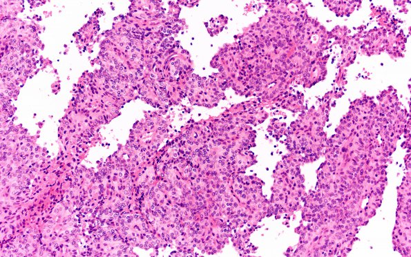
Washington University Experience | NEOPLASMS (MENINGIOMA) | Papillary | 10A2 Meningioma, papillary (Case 10) H&E 20X
Sections show fragments of fibrous tissue and fibrin infiltrated by a papillary meningioma. The neoplasm has cellular, papillary, and nodular growth patterns. The tumor is composed of sheets of hyperchromatic round to oval cells, many of which contain pseudo-nuclear inclusions, suggesting their meningothelial origin but is lacking psammoma bodies. In many locations, the tumor cells line up around blood vessels forming structures resembling perivascular pseudorosettes. The neoplasm has a relatively high mitotic rate but no necrosis is identified.

