Table of Contents
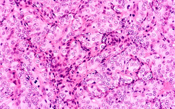
Washington University Experience | NEOPLASMS (MENINGIOMA) | Papillary | 10A5 Meningioma, papillary (Case 10) H&E 40X
The solid appearance has a papillary substructure. ---- Additional data (not shown): Immunohistochemical studies show focal, weak membrane staining with antibodies to EMA. The neoplastic cells are negative for cytokeratin, S100 and type IV collagen by immunohistochemistry. These results are consistent with an atypical meningioma, papillary type, WHO Grade 2.

