Table of Contents
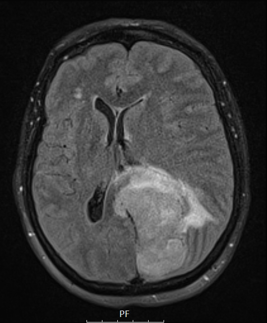
Washington University Experience | NEOPLASMS (MENINGIOMA) | Papillary | 1A1 Meningioma, papillary (Case 1) FLAIR - Copy
Case 1 History ---- The patient was a 51 year-old woman who presented with a six month history of progressive headaches, deterioration of memory and right sided weakness. MRI demonstrated a large (4.4 x 6.6 x 6.1 cm), somewhat heterogeneously-enhancing, T1-isointense, T2-isointense, extra-axial, parasagittal lesion that crossed the midline and was centered on the posterior falx. Cerebral angiography showed the mass was moderately hypervascular with a predominantly pial blood supply. A second, smaller (2.8 x 0.9 x 1.2 cm) extra-axial lesion with similar imaging characteristics was present over the right parietal lobe near the vertex. Radiological diagnosis: Meningioma. Operative procedure: Craniotomy with resection of left parafalcine mass. ---- 1A1-4 MRI Studies: ---- MRI demonstrated a large, FLAIR hyperintense lesion with surrounding edema.

