Table of Contents
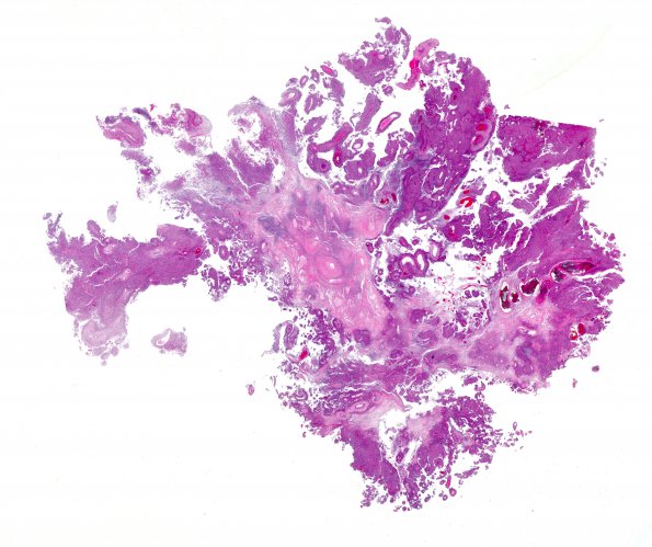
Washington University Experience | NEOPLASMS (MENINGIOMA) | Papillary | 1B1 Meningioma, papillary (Case 1) H&E WM
This H&E stained whole mount shows the papillary appearance of this tumor. The majority of the tumor tissue shows papillary architecture with multiple layers of tumor cells arranged around vascular cores, accompanied by related pseudopapillary architecture and pseudorosettes.

