Table of Contents
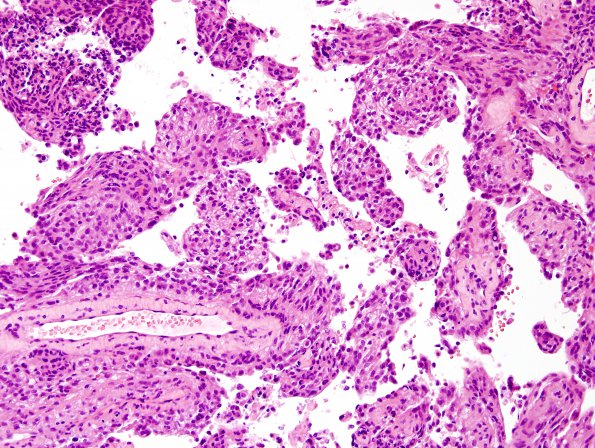
Washington University Experience | NEOPLASMS (MENINGIOMA) | Papillary | 1B4 Meningioma, papillary (Case 1) C6 4.jpg
1B4-7 The tumor cells are generally spindled or epithelioid, with oval nuclei, moderate hyperchromatism, occasional intranuclear cytoplasmic pseudo-inclusions, and a moderate amount of eosinophilic (or occasionally cleared) cytoplasm. Intercellular demarcation is variable, as is nucleolar size. In some areas, the neoplastic cells appear in whorls and fascicles. In other areas, the tumor shows no distinguishing architecture ('sheeting'); other 'atypical' features are also present, including: hypercellularity, 'small cell' change (focally increased tumor cell density, with reduced cytoplasm and increased hyperchromatism), macronucleoli, and spontaneous necrosis. Geographic necrosis is also observed. Rare cells show increased nuclear pleomorphism. Mitotic figures are common and are focally quantified at a density of 7/10HPF. Definitive evidence of brain invasion is not appreciated.

