Table of Contents
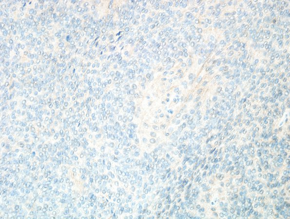
Washington University Experience | NEOPLASMS (MENINGIOMA) | Papillary | 2F Meningioma, papillary (Case 2) CD99 1.jpg
The tumor does not stain for CD99 (CD99 IHC) ---- Ancillary tests (not shown): HMB-45 is negative in tumor cells. Bcl-2 highlights lymphocytes, but tumor cells are negative. ---- FISH showed a deletion of 22q, but normal dosages (2 copies) of chromosome 1, 9 and 14q. This genetic pattern is consistent with a meningioma, but does not show deletions of chromosomal regions commonly encountered in higher grade forms. ---- Comment: The histopathologic, immunohistochemical, and genetic features are consistent with the diagnosis of papillary meningioma, WHO Grade 2. Initially (14 years ago) this tumor was diagnosed as anaplastic meningioma, Grade 3 with the comment that “Without the prominent papillary morphology, this tumor would otherwise qualify for atypical meningioma, but does not have sufficient features for an anaplastic designation. The genetic features of the papillary variant of meningioma have not been thoroughly studied, but it is interesting that in this case, we did not find any of the most common deletions encountered in atypical and anaplastic meningiomas (e.g. 1p, 14q, and 9p). However, it is not clear whether or not in the setting of a papillary meningioma, the lack of these genetic alterations would translate into a less aggressive behavior than typically seen in other WHO grade 3 meningiomas.”

