Table of Contents
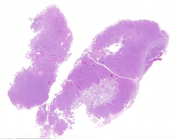
Washington University Experience | NEOPLASMS (MENINGIOMA) | Papillary | 3A1 Meningioma, papillary & rhabdoid (Case 3) 1 H&E WM
Case 3 History ---- The patient was a 61 y/o male who was originally diagnosed with an "unusual meningioma" 11 years prior to his current presentation. This neoplasm was originally located in the temporo-parietal region, and has clinically recurred at that site. ---- Microscopic sections from the 1996 and 2007 revealed an identical tumor, namely a meningioma with rhabdoid and papillary features, WHO grade III. Only small fragments of tissue were available from the 1996 specimen, but abundant neoplastic tissue is available from the 2007 surgery. ---- 3A1,2 The low magnification images of the neurosurgical specimen shows both solid and papillary areas.

