Table of Contents
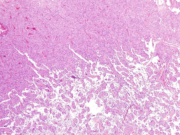
Washington University Experience | NEOPLASMS (MENINGIOMA) | Papillary | 3A6 Meningioma, papillary & rhabdoid (Case 3) H&E 12.jpg
In this image part of the tumor is strongly papillary. The tumor cells involved in the perivascular pseudorosettes have a broad based attachment to their respective blood vessels and accordingly take on a cuboidal to columnar morphology. (H&E)

