Table of Contents
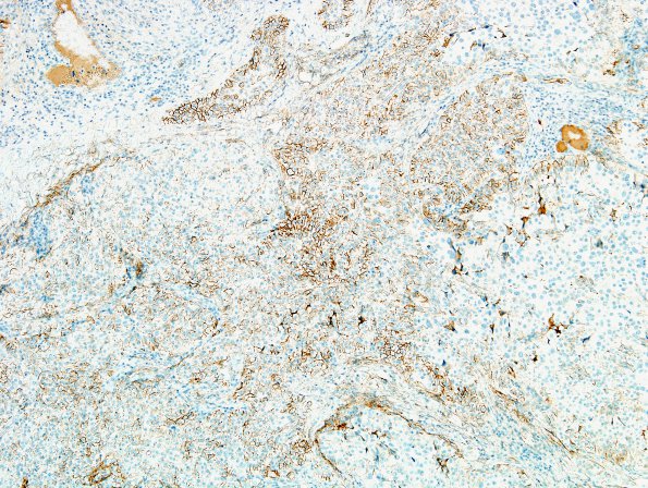
Washington University Experience | NEOPLASMS (MENINGIOMA) | Papillary | 3B1 Meningioma, papillary & rhabdoid (Case 3) minimal EMA 2.jpg
At the margins of solid H&E areas papillary forms are being created from the solid mass, reminding me of the calving of icebergs from glaciers. ---- 3B1-4 Focal membranous EMA positivity is noted but the stain is patchy and weak.

