Table of Contents
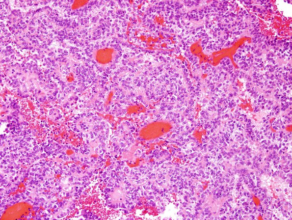
Washington University Experience | NEOPLASMS (MENINGIOMA) | Papillary | 4A1 Meningioma, papillary (Case 4) H&E 1.jpg
Case 4 History ---- The patient was a 19 year old woman with a large, superficial, dural-based lesion. ---- 4A1-5 Sections reveal a predominantly extra-axial papillary neoplasm with moderate to marked hypercellularity. The neoplastic cells have an epithelioid, "carcinoma-like" appearance. They are characterized by round to cuboidal cytology with clear to amphophilic cytoplasm. The papillae are characterized by central vessels surrounded by multi-layered neoplastic lining. In some regions, there is a perivascular nuclear-free zone suggestive of perivascular pseudorosettes. The mitotic index is high with up to 12 mitoses per 10 HPF. The neoplasm has a broad, sharp interface with adjacent brain parenchyma without convincing brain invasion.

