Table of Contents
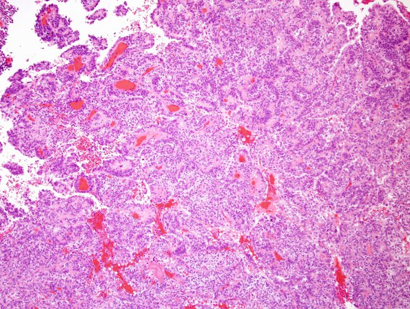
Washington University Experience | NEOPLASMS (MENINGIOMA) | Papillary | 4A2 Meningioma, papillary (Case 4) H&E 2.jpg
Sections reveal a predominantly extra-axial papillary neoplasm with moderate to marked hypercellularity. They are characterized by round to cuboidal cytology with clear to amphophilic cytoplasm. The papillae are characterized by central vessels surrounded by multi-layered neoplastic lining. In some regions, The mitotic index is high with up to 12 mitoses per 10 HPF. The neoplasm has a broad, sharp interface with adjacent brain parenchyma without convincing brain invasion.

