Table of Contents
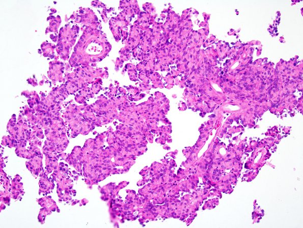
Washington University Experience | NEOPLASMS (MENINGIOMA) | Papillary | 6A1 Meningioma, Papillary (Case 6) H&E 3.jpg
Case 6 History ---- The patient was a 64 year old man with an occipital lobe mass, who had received prior radiation therapy. Operative procedure: Resection. ---- 6A1-4 This is a moderate to highly cellular, relatively solid neoplasm arranged in a predominantly papillary architecture. The tumor cells are relatively small and monomorphic with oval nuclei and small to moderate quantities of eosinophilic cytoplasm. Some of the tumor nuclei have intranuclear pseudoinclusions. The cells are somewhat discohesive and there are frequent perivascular nuclear-free zones resembling perivascular pseudorosettes. Small fragments of brain parenchyma are also seen, although there is no definite evidence of brain invasion. There are scattered mitotic figures reaching 4 mitoses/10HPF. There is abundant tumor necrosis, although it is difficult to determine whether this represents spontaneous necrosis or radiation induced necrosis.

