Table of Contents
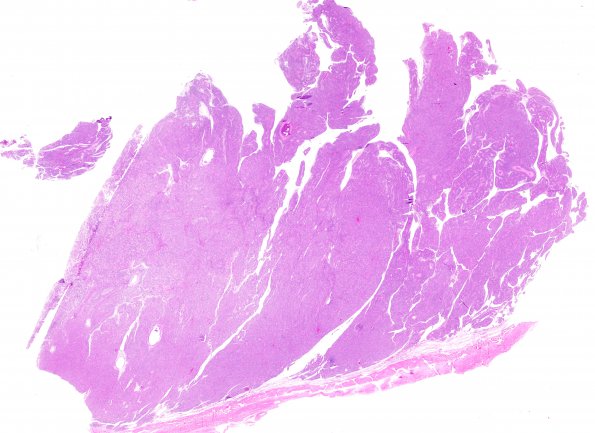
Washington University Experience | NEOPLASMS (MENINGIOMA) | Papillary | 7A1 Meningioma, papillary (Case 7) H&E WM 2
Case 7 History --- The patient was a 58 year old man who has a past history of cerebellar tumor s/p resection in 1964 resulting in shunt placement that has been revised twice most recently in the 1970s. He now presented with ataxia, dizziness and left arm weakness and was found to have a right frontal extra-axial tumor on imaging, s/p resection in 2012. ---- 7A1-9 H&E stained sections from the "right frontal tumor," show a meningothelial neoplasm, arranged in solid sheets. However, intermixed areas show prominent perivascular pseudo-rosetted pattern (akin to ependymoma), consistent with areas of papillary phenotype. The neoplastic cells are rather uniform with moderate to abundant cytoplasm and round nuclei with rare intranuclear cytoplasmic inclusions. Areas of hypercellularity, focal presence of macronucleoli and multiple foci of necrosis are additionally noted. Mitotic figures are scattered focally reaching ~6/10 HPF.

