Table of Contents
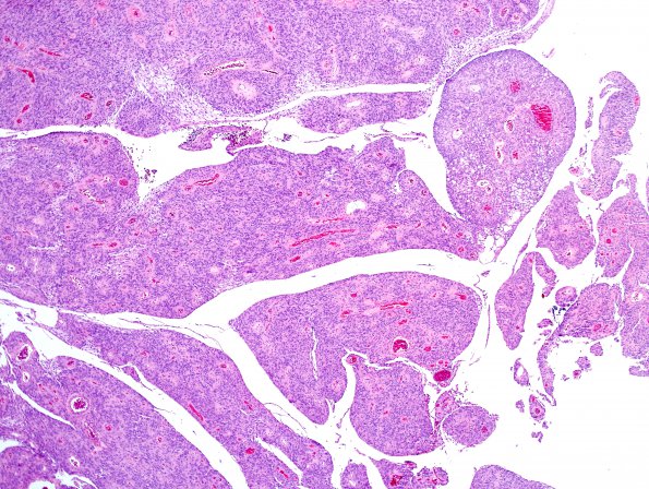
Washington University Experience | NEOPLASMS (MENINGIOMA) | Papillary | 7A3 Meningioma, papillary (Case 7) H&E 11.jpg
H&E stained sections from the "right frontal tumor," show a meningothelial neoplasm, arranged in solid sheets. However, intermixed areas show prominent perivascular pseudo-rosetted pattern (akin to ependymoma), consistent with areas of papillary phenotype.

