Table of Contents
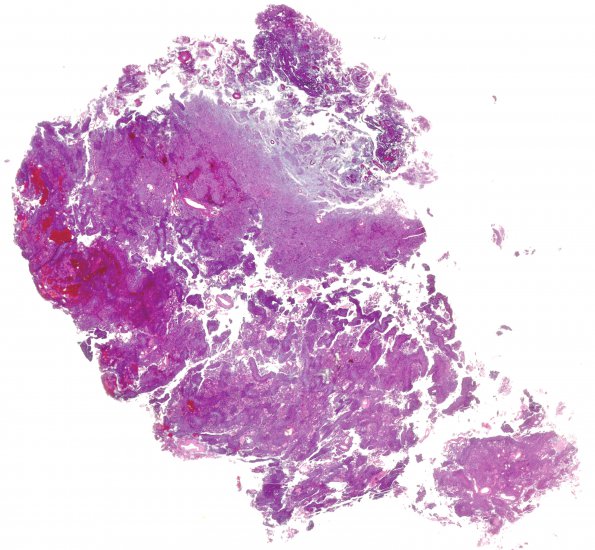
Washington University Experience | NEOPLASMS (MENINGIOMA) | Papillary | 8B1 Meningioma, with papillary features, WHO II (Case 8) H&E WM
8B1-5 Most of the tumor cells are epithelioid or spindled, with oval nuclei, moderate amounts of eosinophilic cytoplasm, and poor intercellular demarcation. Although some areas show occasional classic tight whorls, the majority of the tumor tissue has papillary architecture. Many blood vessels are thickened and hyalinized and bounded by just a few layers of tumor cells, with or without perivascular pseudorosette-like architecture; other papillary structures are about two-dozen cells across, and lack well-defined fibrovascular cores. In addition to this papillary architecture, the tumor shows hypercellularity, lesser non-papillary areas with sheeted architecture, multifocal spontaneous necrosis (<10% of tumor volume), areas with macronucleoli; but foci of small cell change are equivocal. Mitotic figures are uncommon, only focally reaching a density of 2/10HPF.

