Table of Contents
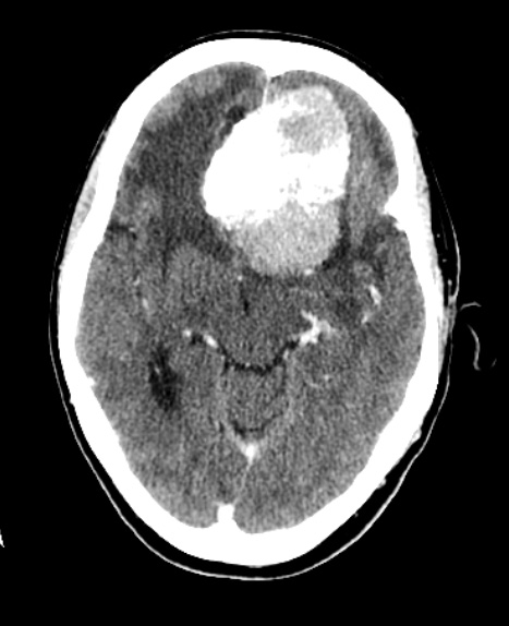
Washington University Experience | NEOPLASMS (MENINGIOMA) | Psammomatous | 1A1 Meningioma, atypical with psamommas (Case 1) CT with Contrast - Copy
Case 1 History ---- The patient is a 67 year-old woman with history of progressive confusion, agitation, falls, left eye blindness, and personality change. Brain MRI showed a large homogeneously enhancing extra-axial dural based mass along the floor of the anterior cranial fossa with surrounding extensive bifrontal vasogenic edema which grows into the sella and suprasellar cistern. Operative procedure: Endoscopic endonasal transsphenoidal skull base approach to the anterior fossa, Stealth frameless stereotaxy, resection of intradural anterior skull base tumor. ---- 1A1 CT There is a hyperdense mass lesion with mineralization and/or hemorrhage.

