Table of Contents
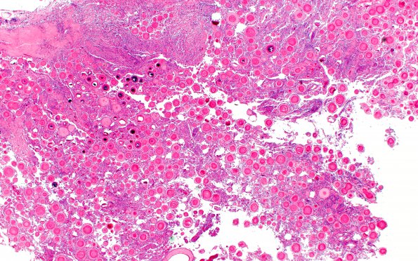
Washington University Experience | NEOPLASMS (MENINGIOMA) | Psammomatous | 1B1 Meningioma, atypical with psamommas (Case 1) H&E 2X
1B1-4 Routine hematoxylin and eosin stained sections show a psammomatous meningioma at different magnifications. The tumor cells intercalated between psammoma bodies have oval, slightly irregular nuclei with scattered intranuclear clearing and pseudoinclusions. There are macronucleoli, focal small cell change, and multiple foci of necrosis. Mitotic activity is focally up to 3/10 high-power fields. Multiple foci of brain invasion are also appreciated.

