Table of Contents
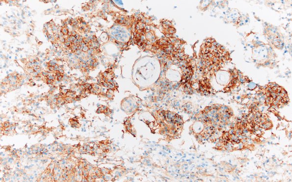
Washington University Experience | NEOPLASMS (MENINGIOMA) | Psammomatous | 1C Meningioma, atypical with psamommas (Case 1) EMA 20X
EMA is focally positive between psammoma bodies. (EMA IHC) ---- Not shown: PR is lost in most tumor cells. GFAP confirms areas of brain invasion. Ki-67 highlights of proliferative index that is focally up to 16.7%. ----- Comment: The overall findings are most consistent with an atypical meningioma, psammomatous variant, with brain invasion, WHO grade II.

