Table of Contents
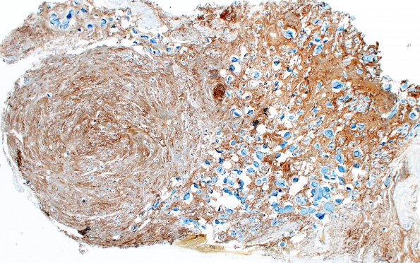
Washington University Experience | NEOPLASMS (MENINGIOMA) | Psammomatous | 3D Meningioma, Hyalinized, psammomatous (Case 3) EMA 2X
Immunohistochemical stains show the tumor cells stain for EMA in this whole mount.---- Not shown: Tumor cells are rarely positive for PR. Ki-67 highlights a proliferation index of less than 1%. ---- Comment: The histopathological and immunohistochemical findings are consistent with meningioma, psammomatous subtype, WHO grade I.

