Table of Contents
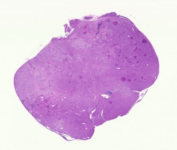
Washington University Experience | NEOPLASMS (MENINGIOMA) | Rhabdoid | 11A1 Meningioma, rhabdoid, Lymph node (Case 11) H&E WM
Case 11 History ---- The patient was a 71 year old woman with history of a T8-T9 meningioma, WHO grade I resected in 2001. Imaging in 2005 and 2006 reportedly showed an enhancing lesion near the T8/9 nerve root, new since 2004. A series of follow-up computed tomography and magnetic resonance imaging studies over the following years showed gradual growth of this lesion, which involved soft tissue and the T8 and T9 vertebrae. PET-CT scan in 1/2013 showed this mass to be hypermetabolic, and also identified slightly hypermetabolic lymph nodes in the neck, axilla, aortocaval area, retroperitoneum and pelvis, as well as a solid right renal mass. In 02/2013, she underwent resection of the "T9 lesion," with pathology reviewed at Mayo Clinic, yielding the diagnosis "Rhabdoid meningioma (WHO grade 3) in progression from a low grade meningioma." In November 2013, she underwent resection of a left neck mass yielding the diagnosis: atypical meningioma (WHO Grade 2) which is re-reviewed as the current case. ---- A whole mount of a neck lymph node overrun by a metastatic anaplastic rhabdoid neoplasm. (H&E)

