Table of Contents
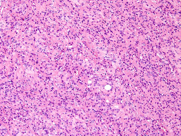
Washington University Experience | NEOPLASMS (MENINGIOMA) | Rhabdoid | 11A2 Meningioma, rhabdoid (Case 11) H&E 2.jpg
11A2-5 Sections show a lymph node expanded by a hypercellular tumor. Most of the tumor cells have round to oval, mildly irregular nuclei, coarsely speckled or vesiculated chromatin, occasional intranuclear pseudoinclusions, modest to moderate eosinophilic cytoplasm, and indistinct cell borders. Some nucleoli appear prominent, but are not clearly visible with a 10x objective. Although weak fascicles can be appreciated, the tumor is generally arranged in a sheet-like pattern. Within this pattern, spontaneous necrosis is noted in several areas, lipomatous metaplasia is regionally abundant, and mitotic activity is high, reaching 22/10HPF. A small fraction of the tumor tissue (approximately 10%) is populated by cells with rhabdoid morphology (eccentric nuclei and large, pale, whorled eosinophilic cytoplasmic inclusions).

