Table of Contents
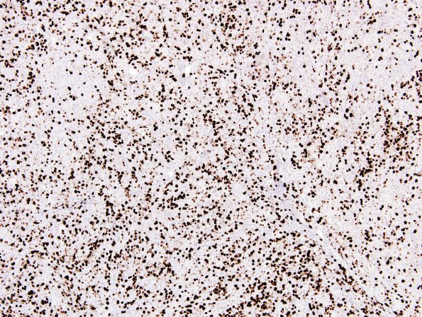
Washington University Experience | NEOPLASMS (MENINGIOMA) | Rhabdoid | 11E Meningioma, rhabdoid, Lymph node (Case 11) Ki67 3.jpg
1E Ki67 immunoreactivty is strikingly increased. ---- Ancillary data (not shown): There are inclusions stained for multiple cytokeratins (AE1/AE3,CAM5.2) and no reactivity for S-100, GFAP, neurofilament, SOX-10, CEA mono, CEA poly, or progesterone receptor. Proliferation marker Ki67 stains approximately 50-60% of tumor cells, and mitotic marker PHH3 stains scattered cells. Immunohistochemistry for CD34 highlights blood vessels only. Factor XIIIa shows positive cytoplasmic reactivity in only a small subset of tumor cells. Reactivity for Bcl-2 highlights residual lymphocytes and only scattered tumor cells. CD99 reactivity is widespread, and weak to moderate. ---- FISH studies performed at WU show evidence for loss of 22q12, and for deletion of the CDKN2A locus at chromosome 9p21. ---- Comment: The morphological, histochemical, immunohistochemical, and genetic features of the left neck mass support the diagnosis 'anaplastic meningioma with focal rhabdoid features, WHO grade 3. This grade is warranted in this case not for the rhabdoid component, but rather, because the mitotic index exceeds the anaplastic threshold of 20/10HPF. Consistent with WHO grade 3 and relatively aggressive behavior, this tumor shows loss of chromosome 9p21. For additional insight into this case, we requested and obtained H&E stained slides from the patient's previous "T9 lesion" resection specimen. By our assessment, the "T9 lesion" is a meningioma with hypercellularity, focal spontaneous necrosis, 'sheeting,' regional lipomatous metaplasia, and a mitotic index that just exceeds 4/10HPF fields; in isolation, these features would support WHO grade 2. Although the previous "T9 lesion" specimen is less cellular than the "left neck mass," the histological patterns share similarities, including rhabdoid features and robust multifocal lipomatous metaplasia. These findings, in conjunction with the history of a thoracic spinal meningioma resected in 2001, support that the "left neck mass" represents a metastasis from the primary "T9 lesion." A diagnosis was made of Metastatic anaplastic meningioma with focal rhabdoid features, WHO Grade 3.

