Table of Contents
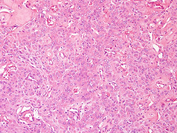
Washington University Experience | NEOPLASMS (MENINGIOMA) | Rhabdoid | 12A1 Meningioma, rhabdoid WHO ungraded (Case 12) H&E 5.jpg
Case 12 History ---- The patient was a 61 year old male with a dural based left fronto-parietal tumor. ---- 12A1-3 Microscopic sections reveal a rhabdoid meningioma. Tumor cells are arranged primarily in patternless sheets within which there is a limited degree of tumor cell loss of cohesion. Tumor cell morphology is predominantly rhabdoid; round to oval nuclei bear an open chromatin pattern and a prominent belly of eosinophilic cytoplasm. Notably, nucleoli are not especially prominent. In addition, mitotic activity is only rare.

