Table of Contents
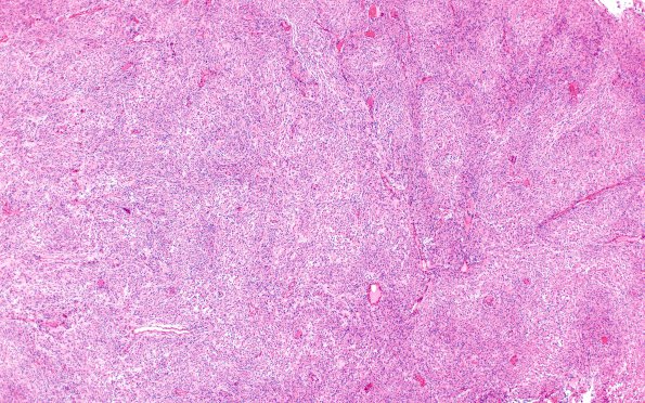
Washington University Experience | NEOPLASMS (MENINGIOMA) | Rhabdoid | 16A1 Rhabdoid Meningioma (Case 16) H&E X4
Case 16 History ---- The patient was a 60 year old woman with a left parietal lesion. Clinical diagnosis: Meningioma. Operative procedure: Craniotomy. ---- 16A1-5 Sections of the left parietal lesion show a dura-associated meningioma composed of atypical meningothelial and bland spindled cells arranged in whorls, bundles and lobules. Atypical features include areas with cellular sheeting, atypical cells with prominent nucleoli, focal necrosis, and rare mitoses. In addition, there are multiple foci of desmin immunonegative tumor cells with rhabdoid features that constitute approximately 5-10% of the tissue.

