Table of Contents
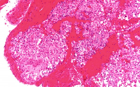
Washington University Experience | NEOPLASMS (MENINGIOMA) | Rhabdoid | 17A2 (Case 17) 20X H&E
Sections of the 3rd ventricle mass show sheets of epithelioid and spindled cells with round to oval, irregular nuclei and scant to abundant eosinophilic cytoplasm, arranged as sheets occasionally forming fascicles or whorls. Some of the cells have abundant eosinophilic cytoplasm with the nucleus pushed toward one side consistent with rhabdoid features. Some cells have prominent nucleoli with occasional intranuclear pseudoinclusions/nuclear clearing. Mitoses are difficult to find and there is no necrosis or vascular proliferation.

