Table of Contents
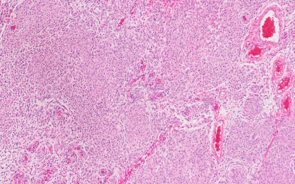
Washington University Experience | NEOPLASMS (MENINGIOMA) | Rhabdoid | 18A1 Meningioma, rhabdoid (Case 18) H&E 10X 2
Case 18 History ---- The patient was a 36 year old man with a large left frontal lobe tumor. ---- 18A1-3 Sections reveal a meningothelial meningioma with focal rhabdoid features. This is manifested by loss of the underlying whorling architecture with "sheeting" and discohesive growth pattern, eccentric nuclei with prominent nucleoli, and abundant eosinophilic cytoplasm with occasional paranuclear inclusions. Small foci of necrosis are also identified. Although the mitotic index is low throughout most of the tumor, five mitoses/10HPF are encountered in one area. Frankly anaplastic features were not found. Based on the focally increased proliferative activity however, an atypical grade (WHO grade 2) is warranted.

