Table of Contents
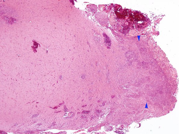
Washington University Experience | NEOPLASMS (MENINGIOMA) | Rhabdoid | 1A1 Meningioma, rhabdoid (Case 1) H&E 2X AP copy.jpg
Case 1 History ---- The patient is a 26 year old man who underwent two resections for different brain tumors. The first surgery in January 2007 was for ganglioglioma (not shown). The second surgery in October 2007 was for a meningioma. Both surgeries were performed at an OSH. ---- 1A1-6 The microscopic appearance of the meningioma specimen varies from intersecting fascicles of relatively bland-appearing spindled cells with scattered whorl formation to large, discohesive sheets of pleomorphic, rhabdoid cells with vesicular nuclei, prominent nucleoli, and eccentric bellies of eosinophilic cytoplasm. The latter component shows a high mitotic index and large zones of tumor necrosis. ---- 1A1 This low magnification image shows the infiltrative margin (arrowhead). (H&E)

