Table of Contents
Washington University Experience | NEOPLASMS (MENINGIOMA) | Rhabdoid | 1A2 Meningioma, rhabdoid (Case 1) H&E 1.jpg
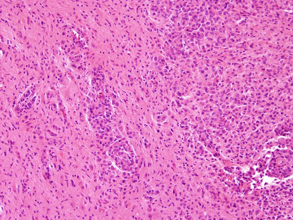
The edge of the infiltration shows growth in the Virchow-Robin spaces and free in the parenchyma. Notice the large number of proliferated hyperplastic microglia (“rod cells”). (H&E)

