Table of Contents
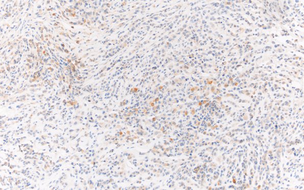
Washington University Experience | NEOPLASMS (MENINGIOMA) | Rhabdoid | 1B Meningioma, rhabdoid (Case 1) EMA 20X 2
EMA histochemistry of the infiltrating tumor shows scattered cells with membranous staining pattern. The EMA stain highlights predominantly the meningothelial-appearing complement of tumor cells, although some of the rhabdoid cells are also positive. (EMA IHC)

