Table of Contents
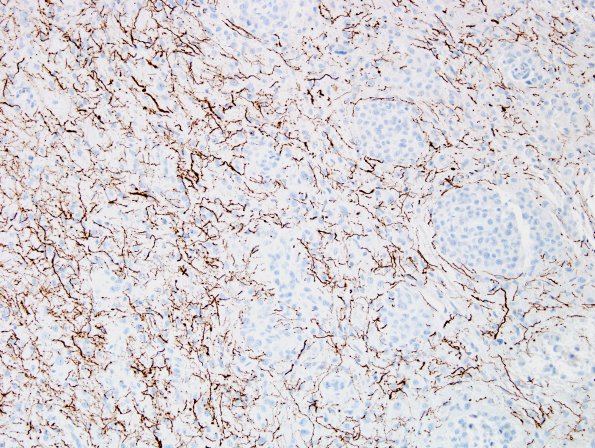
Washington University Experience | NEOPLASMS (MENINGIOMA) | Rhabdoid | 1E Meningioma, rhabdoid (Case 1) NF 4.jpg
Higher magnification images of the infiltrative margin of the tumor showing clumps and individual cells intercalated between glial processes. (GFAP IHC)

