Table of Contents
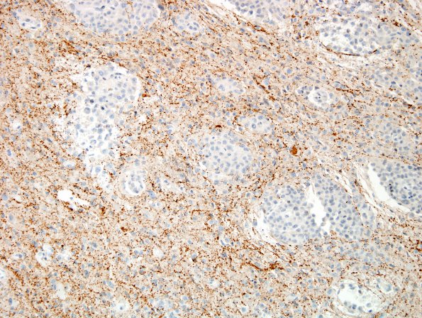
Washington University Experience | NEOPLASMS (MENINGIOMA) | Rhabdoid | 1F Meningioma, rhabdoid (Case 1) SYN 1.jpg
A similar pattern of displaced synaptophysin immunoreactive elements produced by islands of infiltrative tumor. (SYN IHC). ---- Ancillary Findings (not shown): The tumor is reticulin rich. The vimentin stain is diffusely positive and highlights the rhabdoid morphology. The tumor cells are negative for cytokeratin and actin. A stain for INI-1 (BAF47) reveals retained nuclear immunoreactivity within tumor cells, essentially excluding the possibility of an atypical teratoid/rhabdoid tumor. ---- FISH was performed showing polysomy 22 (chromosomal gain), a non-specific finding. ----
Comment: The morphologic and immunohistochemical features were used to diagnose a rhabdoid meningioma, WHO grade III in the (October) resection specimen 15 years ago, but using more modern criteria would be diagnosed as rhabdoid meningioma, WHO Grade 2.

