Table of Contents
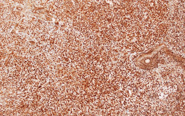
Washington University Experience | NEOPLASMS (MENINGIOMA) | Rhabdoid | 21A (Case 21) VIM 10X
Case 21 History ---- The patient was a 22 year old female with a dural based tumor located in the left parietal region. ---- The microscopic findings are from a report since most slides are no longer available: Microscopic sections reveal an anaplastic meningioma with both clear cell and rhabdoid features, WHO grade 3. Tumor cell density is high in this dural based neoplasm bearing multifocal necrosis. Foci of hyalinization (including blood vessels) are scattered throughout the tumor. Tumor cells display a sheet-like architectural pattern without evidence of better differentiated whorling/fascicular areas. Both clear and rhabdoid cells are scattered throughout; each are associated with mostly bland, rounded nuclei containing nucleoli of variable prominence. Areas of rhabdoid morphology focally have a discohesive quality. Mitoses are rare. A lymphoplasmacytic infiltrate accompanies areas of both acute and more remote hemorrhage. ---- 21A1,2 Vimentin shows a discohesive tumor with prominent cytoplasmic staining. The latter stain helps to highlight the focal rhabdoid morphology. (VIM IHC)

