Table of Contents
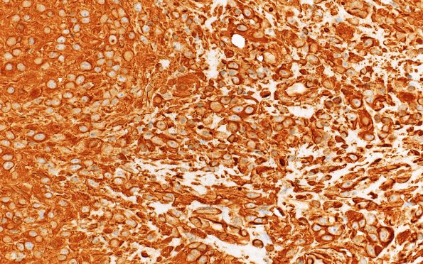
Washington University Experience | NEOPLASMS (MENINGIOMA) | Rhabdoid | 22B (Case 22) VIM 40X
Cells are strongly vimentin immunoreactive (VIM IHC) ---- Ancillary data (not shown): Reticulin staining was performed, and the neoplasm is reticulin rich focally. ---- Immunohistochemical staining with epithelial membrane antigen (EMA), progesterone receptor (PR), glial fibrillary acidic protein (GFAP), Ki-67 (MIB-1), CD99, Bcl-2, and vimentin was performed. EMA and CD99 show patchy positivity. PR is focally positive. Bcl-2 highlights lymphocytes, but tumor cells are negative. The MIB labeling index is markedly elevated, focally reaching up to 40.2%. ---- FISH was performed utilizing probes against BCR, NF2,1p32, 14q23, CEP9, and p16. In this particular case, there were deletions of 22q and 1p, with a subset of tumor cells also showing 14q deletion. There were normal dosages of chromosome 9 with no definite evidence of p16 (chromosome 9p21) deletion. This genetic pattern is consistent with a high-grade meningioma (WHO grades 2 or 3). ---- Comment: The histopathologic, immunohistochemical, and genetic features are consistent with the diagnosis of anaplastic meningioma, WHO Grade 3. Patients with anaplastic meningiomas lacking p16 deletion tend to have somewhat improved overall survival times than those with the deletion. Therefore, this tumor may behave less aggressively than the average anaplastic meningioma.

