Table of Contents
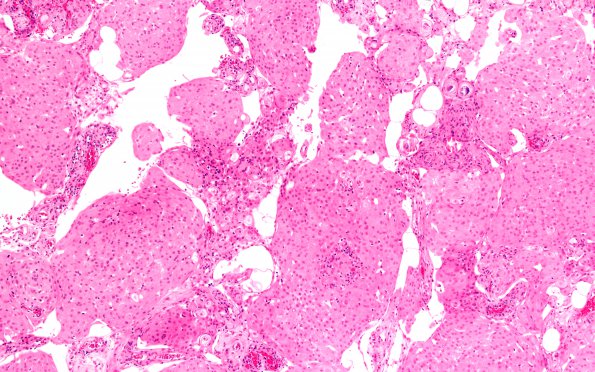
Washington University Experience | NEOPLASMS (MENINGIOMA) | Rhabdoid | 24A2 Rhabdoid meningioma (Case 24) H&E areaA 10X
24A2-4 This is a meningothelial neoplasm arranged predominantly in whorls and a fascicular pattern with small foci of sheeting. The tumor cells are spindled to polygonal, have moderate to abundant eosinophilic cytoplasm and vesicular nuclei, however, focally they do contain prominent nucleoli. In some areas, the tumor cells appear rounder with eccentrically placed nuclei, and have globular clumps of eosinophilic cytoplasm (rhabdoid differentiation). However, these rhabdoid areas do not seem to be fully developed as they lack prominent macronucleoli as well as other obvious malignant features. Patchy areas of hypercellularity with small cell features are also notable. In addition, small foci of brain invasion are identified, that are furthermore highlighted by GFAP immunostain. Mitoses are rare and scattered (1- 2/10hpf) and necrosis is not seen.

