Table of Contents
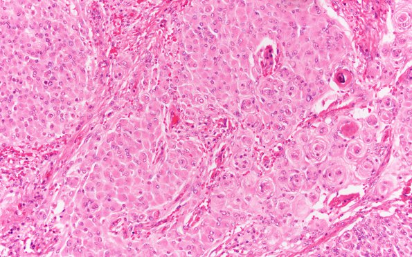
Washington University Experience | NEOPLASMS (MENINGIOMA) | Rhabdoid | 26A1 Meningioma, rhabdoid (Case 26) H&E 20X 3
Case 26 History ---- The patient was a 31 year old woman with an extraaxial, enhancing mass overlying the right frontal and temporal lobes. Operative procedure: Craniotomy ---- 26A1-4 Sections of the material from the extra-axial right frontotemporal region show a heterogeneous meningothelial neoplasm that arises from the dura and is composed of meningothelial, transitional, and atypical components, the latter of which includes focal areas of small cell change, sheeting, and atypical cytology with focal prominent nucleoli. A significant component shows rhabdoid morphology and is composed of oval to elongated cells with abundant, eccentrically placed eosinophilic cytoplasm. Xanthomatous metaplasia and areas of micronecrosis are also apparent. Despite the atypical and rhabdoid features, mitoses are hard to find. Overall, these features are consistent with an atypical meningioma, WHO grade 2, with focal rhabdoid features.

