Table of Contents
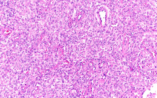
Washington University Experience | NEOPLASMS (MENINGIOMA) | Rhabdoid | 28A1 (Case 28) Poor Quality H&E 20X
Case 28 History ---- The patient was a 63 year old man who initially underwent a meningioma resection in 1999. A recurrence was resected in 2003 and surgery was performed in 2006 for multiple meningioma nodules near the prior site of resection. ---- 28A1,2 Sections reveal a hypercellular dural-based neoplasm. The tumor cells are arranged in fascicles and vague whorls and are composed of medium sized to large cells with oval nuclei, prominent nucleoli, and abundant eosinophilic cytoplasm. In some areas, the cells are arranged in 2 dimensional sheets. There is no definite necrosis or small cell formation. However, there are several foci suspicious for brain invasion. Additionally, there are small foci of rhabdoid morphology with discohesive growth pattern and accumulations of eccentric eosinophilic cytoplasm. Scattered mitotic figures are seen, reaching up to 2 to 3 mitoses per 10 high power fields.

