Table of Contents
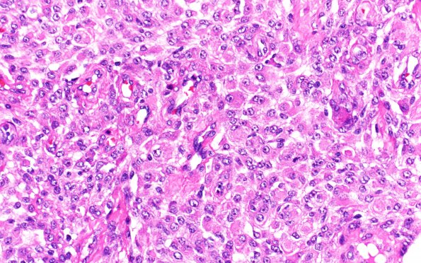
Washington University Experience | NEOPLASMS (MENINGIOMA) | Rhabdoid | 28A2 (Case 28)) Poor Quality H&E 40X
Additional tumor cell image. (H&E) ---- Ancillary findings (not shown): Immunoperoxidase stains were performed showing patchy tumor immunoreactivity for EMA and PR. A stain for GFAP highlights entrapped glial tissue at the periphery of the tumor, consistent with brain invasion. The MIB-1 (Ki-67) labeling index is moderate, average roughly 5.6%. The morphologic and immunohistochemical features are consistent with the diagnosis of atypical meningioma with focal rhabdoid features and brain invasion, WHO grade 2

