Table of Contents
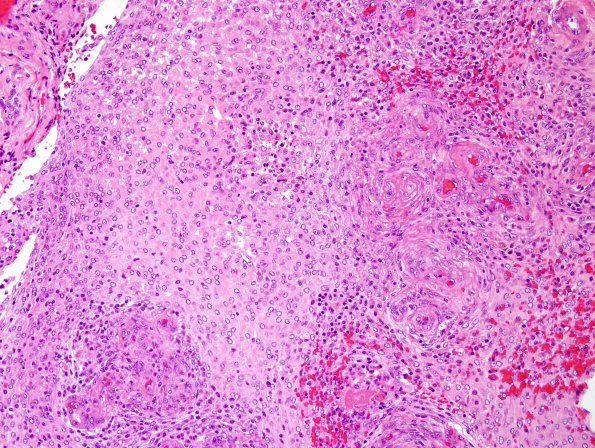
Washington University Experience | NEOPLASMS (MENINGIOMA) | Rhabdoid | 3B2 Meningioma, atypical w rhabdoid features (Case 3) H&E 2.jpg
3B2-6 H&E showed a meningioma with heterogeneous histological patterns. Among areas with angiomatous and transitional features are many sheeted lobules with rhabdoid cytology, i.e., epithelioid cells with eccentric, eosinophilic, finely fibrillar whorled inclusions and round-to-oval nuclei with open coarsely-speckled chromatin involving a slight majority of the tumor volume. Occasionally within these sheeted areas, the tumor vasculature is strikingly hyperplastic and garlanded/glomeruloid. Rare psammoma bodies are noted. The tumor is hypercellular in many areas, and exhibits foci of ‘small cell change,’ spontaneous necrosis (far less than 10% of tumor volume), as well as ‘sheeted’ architecture. Macronucleoli are not appreciated. Mitotic figures are present at generally low density, ranging only focally up to 3/10HPF.

